Optical Coherence Tomography

Optical Coherence Tomography Oct Carabin Eye Care
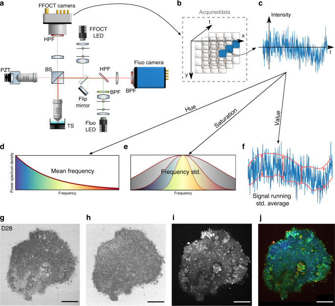
Dynamic Full Field Optical Coherence Tomography 3d Live Imaging Of Retinal Organoids Light Science Applications
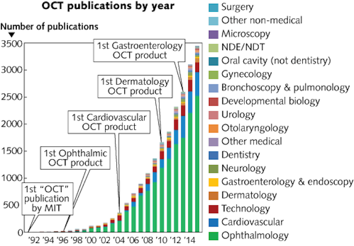
Optical Coherence Tomography Beyond Better Clinical Care Oct S Economic Impact Laser Focus World

The Future Of Optical Coherence Tomography Oct

Optical Coherence Tomography A Review Of The Opportunities And Challenges For Postharvest Quality Evaluation Sciencedirect
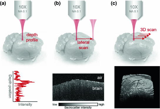
Optical Coherence Tomography For Brain Imaging Springerlink
The use of Optical coherence Tomography (OCT) was essential to solve this problem and determine the modality of treatment The use of DES for re stenting the stent fracture segment was an important decision, the foundation of which laid on the use of OCT, thus improving the mortality, morbidity and prognosis of our patient.

Optical coherence tomography. Optical coherence tomography (OCT) has been used to image anterior and posterior structures of the eye, but it has not been used to image structures behind the sclera 1,2 Extraocular muscles. Optical Coherence Tomography Scans This guide was created to provide information to students, residents and clinicians in eye care to better understand optical coherence tomography scans Examples are provided to help understand optic nerve and retinal pathologies to assist in the diagnosis and management of these conditions. Optical coherence tomography (OCT) is a procedure used for the noninvasive examination of intraocular structures Primarily used for analysis of the retina and optic nerve, OCT centers on the amount of light absorption or scattering that occurs when light passes through a given tissue layer Optical coherence tomography uses a diode laser, which emits light at a wavelength of about 840 nanometers.
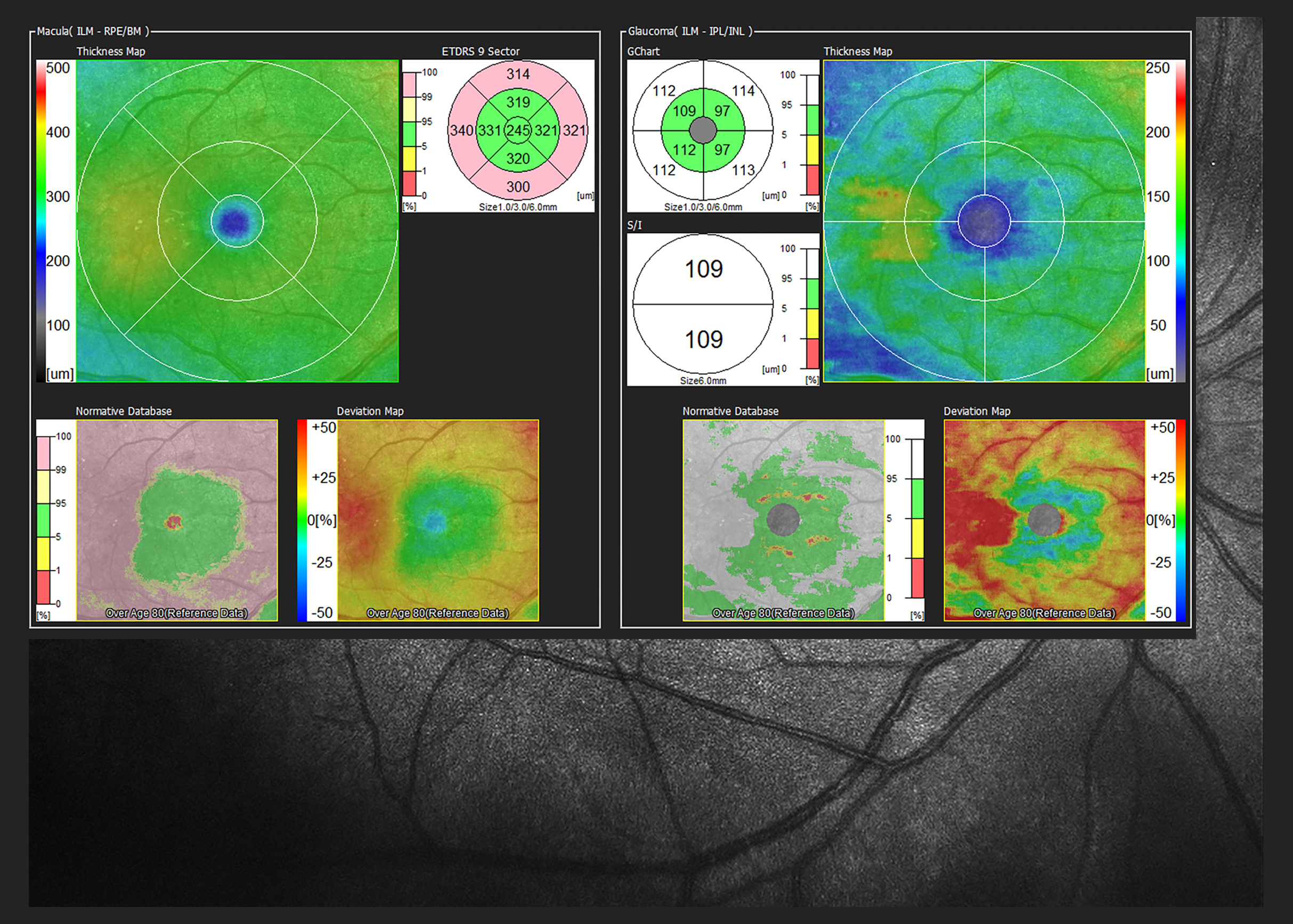
Optical coherence tomography glaucoma progression Because of its objective nature and low test–retest variability, structural SDOCT is a valuable tool to estimate disease progression 39 It is commonly believed that structural OCT is useful only in the preperimetric and early stages of the disease, while it is of limited help in advanced glaucoma. Optical Coherence Tomography (OCT) uses coherent interferometry to construct a crosssectional view of ocular structures that is accurate to less than 10 microns Interferometry is the general technique of superimposing, or "interfering" two or more light waves which creates an output wave that is different from the input waves. Optical Coherence Tomography (OCT) is a noncontact, noninvasive, advanced imaging technique used to obtain highresolution crosssectional images of the retina.
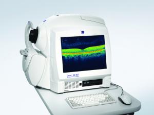
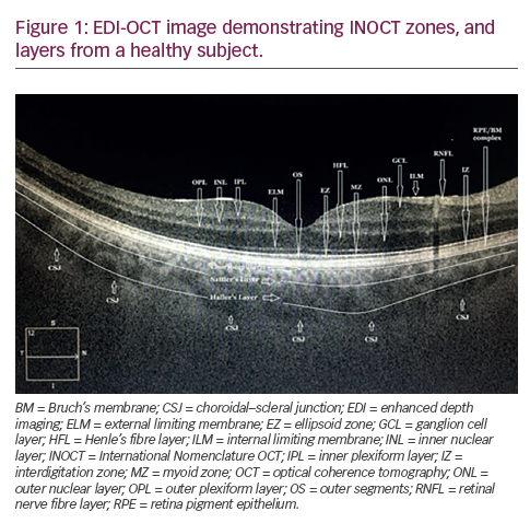
Optical coherence tomography is a way for optometrists and ophthalmologists to image the back of the eye including the macula, optic nerve, retina, and choroid During an eye examination, optometrists and ophthalmologist can view the back of the eye and its anatomy However, sometimes doctors need more detail or need to inspect detail right below the surface which is difficult to view with standard techniques. An optical coherence tomography scan (commonly referred to as an OCT scan) helps us to view the health of your eyes in greater detail, by allowing us to see what’s going on beneath the surface of the eye. Optical coherence tomography (OCT) has been used to image anterior and posterior structures of the eye, but it has not been used to image structures behind the sclera 1,2 Extraocular muscles.
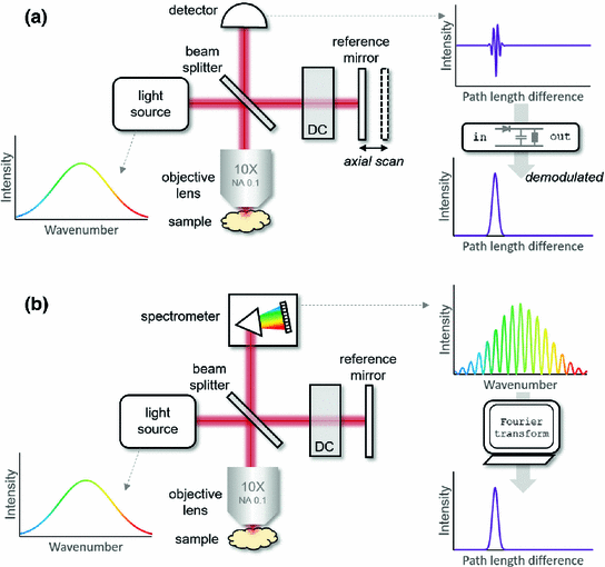
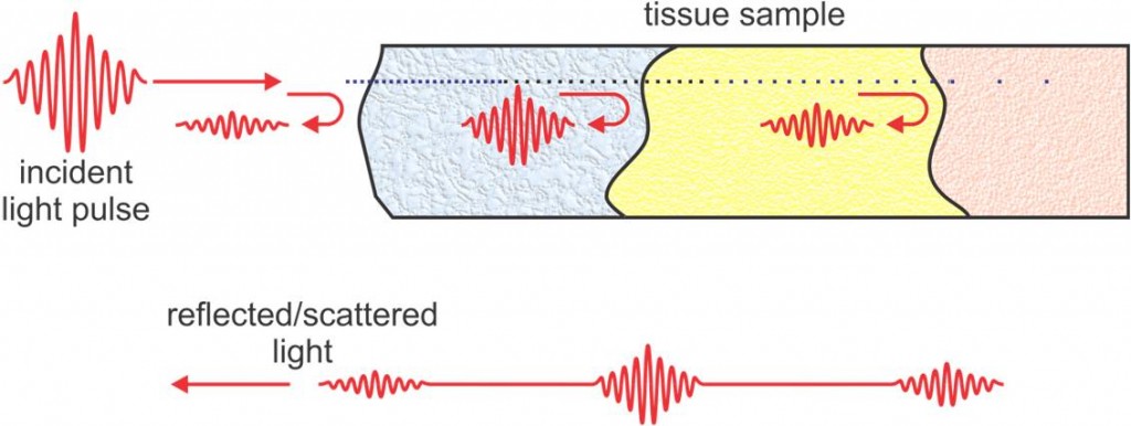
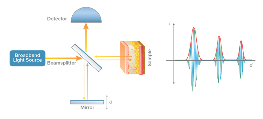
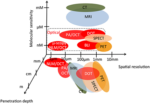
Optical coherence tomography (OCT) 1,2 is an imaging technique which works similar to ultrasound, simply using light waves instead of sound waves By using the timedelay information contained in the light waves which have been reflected from different depths inside a sample, an OCT system can reconstruct a depthprofile of the sample structure. Optical coherence tomography (OCT) is a micronscale imaging method, analogous to ultrasound measuring the backreflection of infrared light rather than sound It can operate in 2D and 3D mode and exceeds the frame rate of video The penetration depth of OCT is about 2 mm, depending on tissue types. Optical coherence tomography (OCT) is a diagnostic procedure that is used during cardiac catheterization Unlike ultrasound, which uses sound waves to produce an image of the blood vessels, OCT uses light With OCT, doctors can obtain images of the blood vessels that are about the same as if they were looking under a microscope.
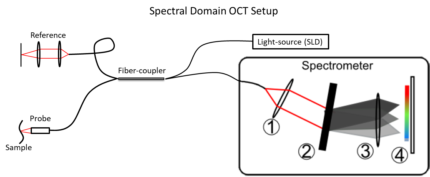
Optical coherence tomography angiography (OCTA) has emerged as a noninvasive technique for imaging the microvasculature of the retina and the choroid The first clinical studies using this innovative technology were published in 14 1. Thorlabs provides solutions for the field of Optical Coherence Tomography (OCT) imaging on the system, subsystem, and component level Our drive for innovation is shaping our entire rapidly expanding product line Complete spectraldomain, swept source, and polarizationsensitive OCT systems are available that are outofthebox ready for biological, industrial, and research applications. Optical coherence tomography (OCT) is an emerging imaging technique for a wide range of biological, medical, and material investigations OCT was initially developed for imaging biological tissue because it permits the imaging of tissue microstructure in situ , yielding micronscale image resolution without the need for excision of a specimen and tissue processing.
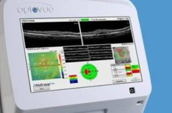
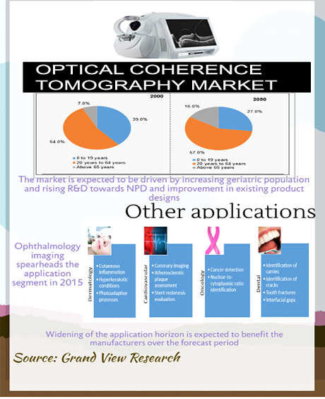
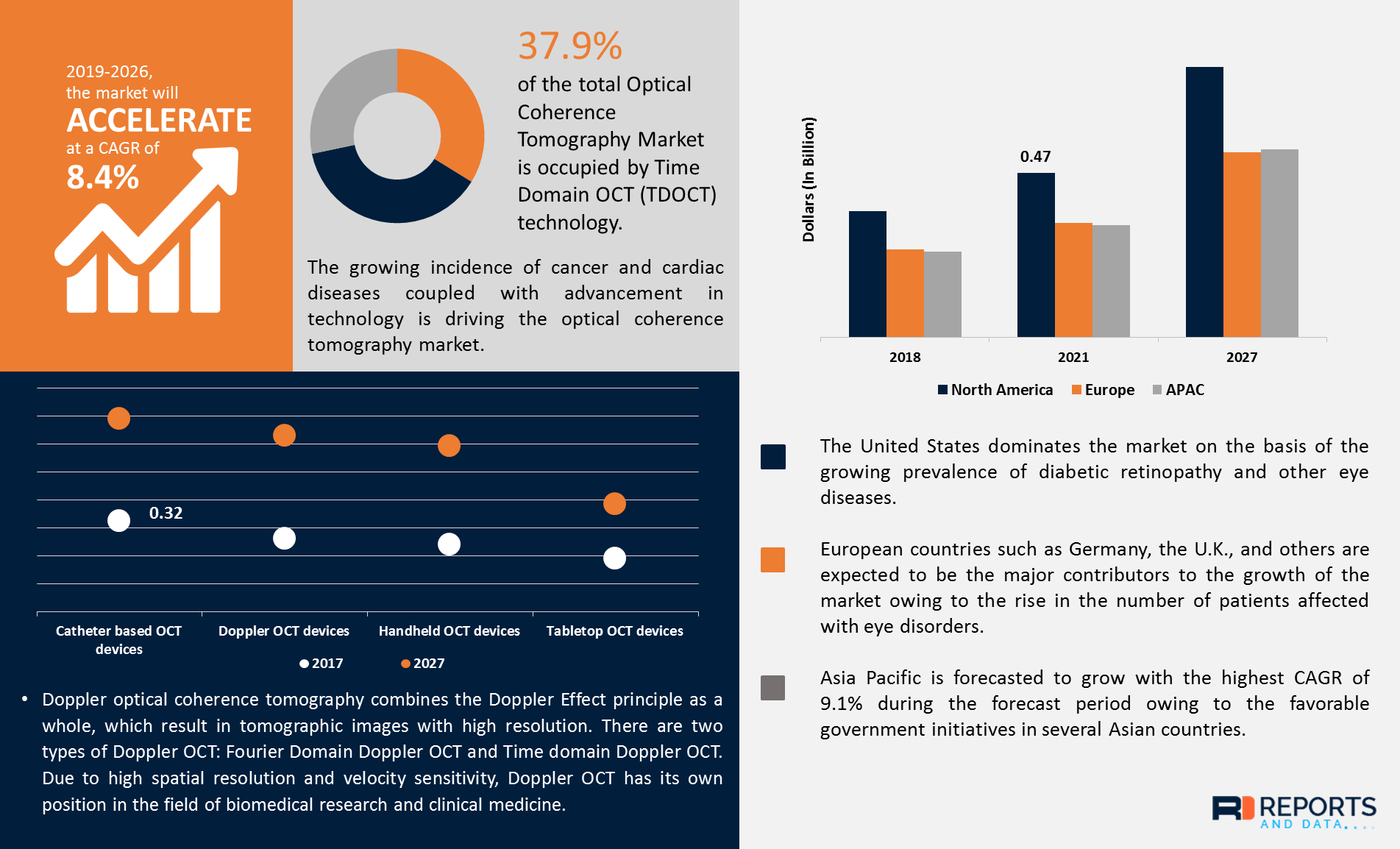
The competitive landscape of the Optical Coherence Tomography market is defined by companies such as The major players covered in Optical Coherence Tomography are,Carl Zeiss Meditec AG,Optovue,Heidelberg Engineering GmbH,Agfa Healthcare,Novacam Technologies Inc,Imalux Corporation,Thorlabs,Michelson Diagnostics,OPTOPOL Technology SA. Optical coherence tomography angiography relies on motion for contrast and requires at least two data acquisitions per pointwise scanning location We present a method termed spectral contrast optical coherence tomography angiography using visible light that relies on the spectral signatures of blood for angiography from a single scan using. The competitive landscape of the Optical Coherence Tomography market is defined by companies such as The major players covered in Optical Coherence Tomography are,Carl Zeiss Meditec AG,Optovue,Heidelberg Engineering GmbH,Agfa Healthcare,Novacam Technologies Inc,Imalux Corporation,Thorlabs,Michelson Diagnostics,OPTOPOL Technology SA.
INTRODUCTION Optical coherence tomography, or OCT is a non contact, noninvasive imaging technique used to obtain high resolution 10 cross sectional images of the retina and anterior segment Reflected light is used instead of sound waves Infrared ray of 0 nm with 78D internal lens 4. Optical coherence tomography (OCT) is a wellestablished noninvasive, 3D imaging technique that has been used in medical application as a diagnostic tool 107 It is based on the analyses of light interference properties generated by nearinfrared light which is split into and recombined from a reference and sample arm. To better comprehend these opaque procedures, a convolutional neural network for optical coherence tomography image segmentation was enhanced with a Traceable Relevance Explainability (TREX.
Optical coherence tomography angiography relies on motion for contrast and requires at least two data acquisitions per pointwise scanning location We present a method termed spectral contrast optical coherence tomography angiography using visible light that relies on the spectral signatures of blood for angiography from a single scan using. Optical coherence tomography is a type of ophthalmic research with the help of which the doctor gets the scans of optically transparent eye tissues The obtained images are characterized by high indices of spatial resolution Types of Optical coherence tomographs A coherence tomograph resembles an ultrasound machine by its operation principle. The Optical Coherence Tomography (OCT) for Ophthalmology market in the US is estimated at US$1924 Million in the year China, the world`s second largest economy, is forecast to reach a.
To better comprehend these opaque procedures, a convolutional neural network for optical coherence tomography image segmentation was enhanced with a Traceable Relevance Explainability (TREX. Optical Coherence Tomography, or otherwise known as an OCT, is a new imaging technique that provides high resolution and crosssectional images of the eye It gives our optician a very unique look at the retina and a real accurate representation of your eye health. Optical coherence tomography is a way for optometrists and ophthalmologists to image the back of the eye including the macula, optic nerve, retina, and choroid During an eye examination, optometrists and ophthalmologist can view the back of the eye and its anatomy However, sometimes doctors need more detail or need to inspect detail right below the surface which is difficult to view with standard techniques.
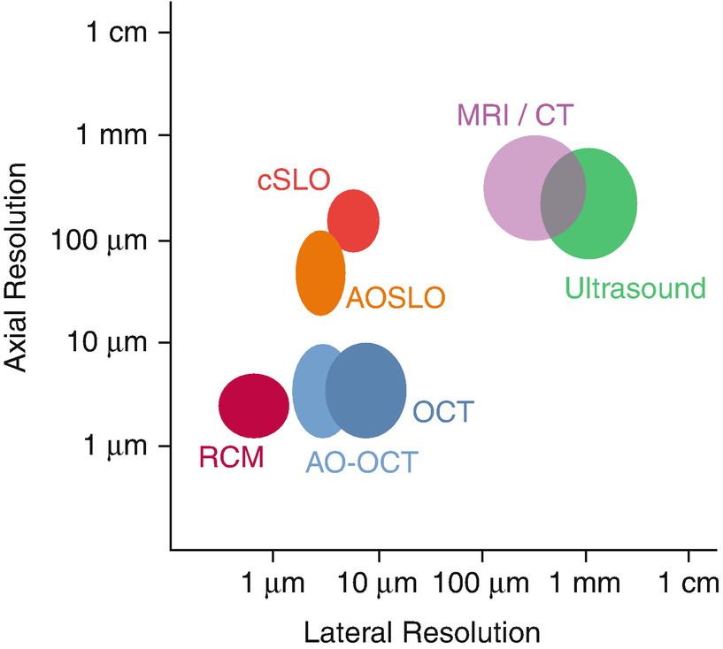
Optical Coherence Tomography is a powerful noninvasive imaging modality that performs high resolution, micronscale, crosssectional imaging of the retina Originally developed in 1991 by Huang et al, 1 OCT technology has continually evolved and expanded within ophthalmologyand has been explored in a wide range of clinical applications. Optical coherence tomography (OCT) is a 3D imaging technique that can provide high resolution imaging in a scattering media, nondestructively and without the need for contact or a coupling medium Lateral imaging resolution on the order of a few micrometers is possible, to depths up to few millimeters. Optical coherence tomography (OCT) 1,2 is an imaging technique which works similar to ultrasound, simply using light waves instead of sound waves By using the timedelay information contained in the light waves which have been reflected from different depths inside a sample, an OCT system can reconstruct a depthprofile of the sample structure.
Optical coherence tomography (OCT) is a relatively new noninvasive imaging modality that uses reflected light in a manner analogous to the use of sound waves in ultrasonography to create highresolution (10 micron) crosssectional images of the vitreoretinal interface, retina and subretinal space, analogous to histological sections seen. Optical coherence tomography (OCT) is a 3D imaging technique that can provide high resolution imaging in a scattering media, nondestructively and without the need for contact or a coupling medium Lateral imaging resolution on the order of a few micrometers is possible, to depths up to few millimeters. Optical coherence tomography(OCT) is a noninvasive imaging technology Utilized to obtain highresolution crosssectional Pictures of the retina to capture micrometerresolution, two and threedimensional Pictures from Inside optical scattering media.
To better comprehend these opaque procedures, a convolutional neural network for optical coherence tomography image segmentation was enhanced with a Traceable Relevance Explainability (TREX. Swept Source OCT DRI OCT Triton Optical Coherence Tomography 3D OCT1 (TypeMaestro2) Smart Infrastructure TOPCON Brand What's new;. Working Principles of OCT Optical coherence tomography has some similarity to laser microscopy (although in many cases a laser source is not used) In two transverse dimensions, image resolution is obtained by scanning a tightly focused light beam over the sample and performing measurements on backscattered light.
Optical Coherence Tomography (OCT) is a noninvasive diagnostic technique that renders an in vivo cross sectional view of the retina OCT utilizes a concept known as inferometry to create a crosssectional map of the retina that is accurate to within at least 1015 microns. Optical coherence tomography angiography (OCTA) has emerged as a noninvasive technique for imaging the microvasculature of the retina and the choroid The first clinical studies using this innovative technology were published in 14 1. Optical coherence tomography (OCT) is a noninvasive imaging test that uses light waves to take crosssection pictures of your retina, the lightsensitive tissue lining the back of the eye My DashboardMy EducationFind an Ophthalmologist.
Optical coherence tomography – or OCT – is a medical imaging technique It uses visible light to produce a 3D image of what’s happening beneath the surface MRI, CTscans and Xrays use radiation for this But this can pose health risks, especially if repeated OCT is more like ultrasound It only uses visible light (rather than sound). Optical coherence tomography (OCT) is a noninvasive diagnostic technique providing crosssectional images of biologic structures based on the differences in tissue optical properties. Optical coherence tomography is a type of ophthalmic research with the help of which the doctor gets the scans of optically transparent eye tissues The obtained images are characterized by high indices of spatial resolution Types of Optical coherence tomographs.
A technique called optical coherence tomography (OCT) has been developed for noninvasive crosssectional imaging in biological systems OCT uses lowcoherence interferometry to produce a twodimensional image of optical scattering from internal tissue microstructures in a way that is analogous to ultrasonic pulseecho imaging. To better comprehend these opaque procedures, a convolutional neural network for optical coherence tomography image segmentation was enhanced with a Traceable Relevance Explainability (TREX. Optical coherence tomography (OCT) is an imaging technique that uses lowcoherence light to capture micrometer resolution, two and threedimensional images from within optical scattering media (eg, biological tissue) It is used for medical imaging and industrial nondestructive testing (NDT).
Light waves near infrared range (1,300 nm wavelength) are projected around the imaged structure and the reflected backscattered light signals form the images for analysis Optical coherence tomography has become established as an imaging modality in clinical ophthalmology and is finding applications in even gastrointestinal and dermatological arenas. Optical coherence tomography (OCT) is an established medical imaging technique that uses light to capture micrometerresolution, threedimensional images from within optical scattering media (eg, biological tissue) Optical coherence tomography is based on lowcoherence interferometry, typically employing nearinfrared light. Optical coherence tomography (OCT) is a 3D imaging technique that can provide high resolution imaging in a scattering media, nondestructively and without the need for contact or a coupling medium Lateral imaging resolution on the order of a few micrometers is possible, to depths up to few millimeters.
Optical coherence tomography (OCT) has become one of the most important techniques in ophthalmic diagnostics, as it is the only way to threedimensionally visualize morphological changes in the layered structure of the retina at a high resolution In addition, OCT is applied for countless medical and technical purposes. Optical Coherence Tomography A test used to follow patients diagnosed with MS on their recovery and response to treatments. Optical Coherence Tomography (OCT) is an imaging technique that provides high resolution, nondestructive, in situ, realtime reflectivity profiling of nonabsorptive samples Medical applications include noninvasive ophthalmological imaging and endoscopic gastrointestinal tract imaging.
Optical coherence tomography ( OCT ) is an imaging technology with applications in biology, medicine, and materials investigations In a clinical environment, OCT has revolutionized the clinical practice of ophthalmology by providing 3D anatomic information. The use of Optical coherence Tomography (OCT) was essential to solve this problem and determine the modality of treatment The use of DES for re stenting the stent fracture segment was an important decision, the foundation of which laid on the use of OCT, thus improving the mortality, morbidity and prognosis of our patient. The competitive landscape of the Optical Coherence Tomography market is defined by companies such as The major players covered in Optical Coherence Tomography are,Carl Zeiss Meditec AG,Optovue,Heidelberg Engineering GmbH,Agfa Healthcare,Novacam Technologies Inc,Imalux Corporation,Thorlabs,Michelson Diagnostics,OPTOPOL Technology SA.
Optical coherence tomography (OCT) has been used to image anterior and posterior structures of the eye, but it has not been used to image structures behind the sclera 1,2 Extraocular muscles. OCT stands for Optical Coherence Tomography, which is a piece of diagnostic equipment that takes a series of advanced 3D scans of the back of the eye These 3D scans of the optic nerve, retina and macula are highly detailed and allow your Specsavers optometrist to view the granular structures of your eye, allowing for a more advanced and accurate examination of your eye health. INTRODUCTION Optical coherence tomography, or OCT is a non contact, noninvasive imaging technique used to obtain high resolution 10 cross sectional images of the retina and anterior segment Reflected light is used instead of sound waves Infrared ray of 0 nm with 78D internal lens.
The competitive landscape of the Optical Coherence Tomography market is defined by companies such as The major players covered in Optical Coherence Tomography are,Carl Zeiss Meditec AG,Optovue,Heidelberg Engineering GmbH,Agfa Healthcare,Novacam Technologies Inc,Imalux Corporation,Thorlabs,Michelson Diagnostics,OPTOPOL Technology SA. The use of Optical coherence Tomography (OCT) was essential to solve this problem and determine the modality of treatment The use of DES for re stenting the stent fracture segment was an important decision, the foundation of which laid on the use of OCT, thus improving the mortality, morbidity and prognosis of our patient. The use of Optical coherence Tomography (OCT) was essential to solve this problem and determine the modality of treatment The use of DES for re stenting the stent fracture segment was an important decision, the foundation of which laid on the use of OCT, thus improving the mortality, morbidity and prognosis of our patient.

Optical Coherence Tomography Market 19 Size Share Current Trends Top Key Companies And Future Insights Forecast To 23 Medgadget

What Is Oct And How Can It Help Ophthalmologists Acquire High Resolution Information On Ocular Tissue Learn Share Leica Microsystems
Q Tbn And9gctjenkjznj4ttj0 I7a8nheky7hx Txzzgmvmxy C4sixfznf31 Usqp Cau

Biomolecular Contrast Agents For Optical Coherence Tomography Biorxiv

Optical Coherence Tomography Oct Left Volumetric Oct Data Shown Download Scientific Diagram

Optical Coherence Tomography Wikipedia The Free Encyclopedia Optical Coherence Tomography Quantum World Optical

Introduction To Oct Obel
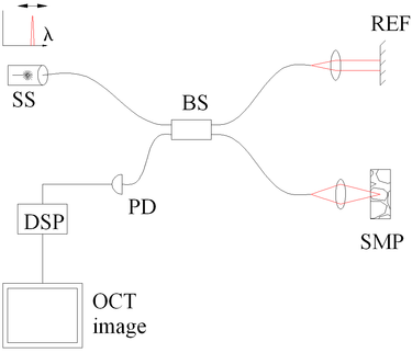
Optical Coherence Tomography Wikipedia

Optical Coherence Tomography Applications Expand As Technology Matures Youtube

Global Optical Coherence Tomography For Ophthalmology Market 24 Evolving Opportunities With Abbott Laboratories And Canon Inc Technavio Business Wire

Advances In Optical Coherence Tomography Features Oct 18 Photonics Spectra

Optical Coherence Tomography Biophotonics Imaging Laboratory
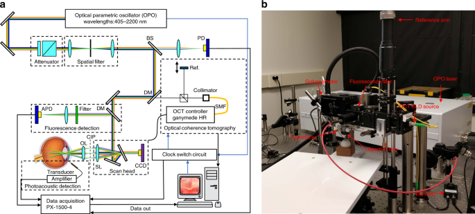
High Resolution In Vivo Multimodal Photoacoustic Microscopy Optical Coherence Tomography And Fluorescence Microscopy Imaging Of Rabbit Retinal Neovascularization Light Science Applications
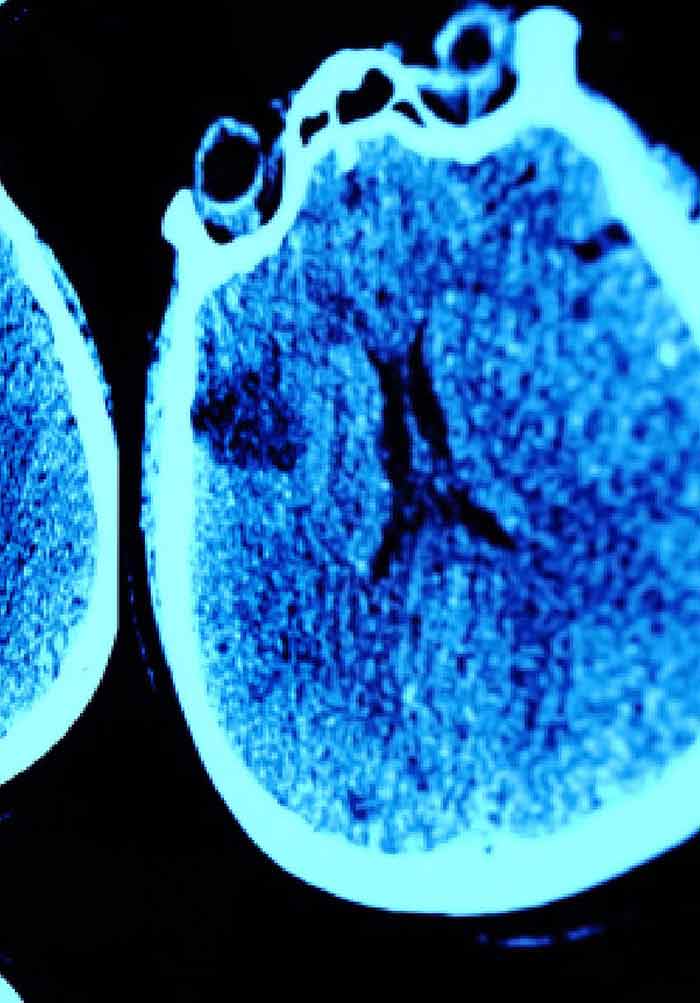
Optical Coherence Tomography Intechopen

Optical Coherence Tomography Wikipedia

What Is Oct Scanning Optical Coherence Tomography Youtube
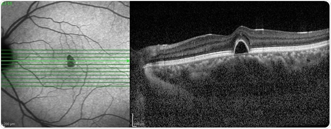
What Is Optical Coherence Tomography

Introduction To Oct Obel
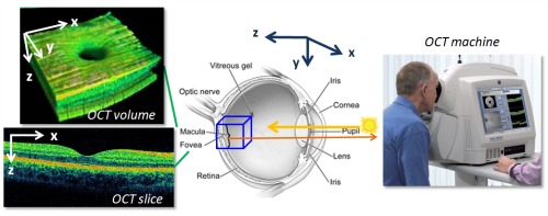
Optical Coherence Tomography Oct Bay St Eyecare

Optical Coherence Tomography Wikipedia

Optical Coherence Tomography Enables More Accurate Detection Of Functionally Significant Intermediate Non Left Main Coronary Artery Stenoses Than Intravascular Ultrasound A Meta Analysis Of 6919 Patients And 7537 Lesions International Journal Of
What Is An Oct Eye Exam

Questioning Optical Coherence Tomography Ophthalmology
Optical Coherence Tomography For Brain Imaging Springerlink

Zeiss Optical Coherence Tomography Bonavista Eye
Optical Coherence Tomography Oct Optical Cancer Imaging Laboptical Cancer Imaging Lab
Optical Coherence Tomography Oct Exam Optometrist Elko Nv

Atlas Of Retinal Oct Optical Coherence Tomography Abdo College

What Is Oct And How Can It Help Ophthalmologists Acquire High Resolution Information On Ocular Tissue Learn Share Leica Microsystems
Q Tbn And9gcqqnsqyzmp8sj0ux Y7rtdsgbv2uk1wlcrr2xc1gnuxipqt5sc1 Usqp Cau
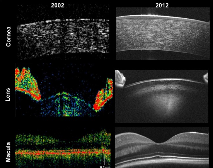
Twenty Five Years Of Optical Coherence Tomography The Paradigm Shift In Sensitivity And Speed Provided By Fourier Domain Oct Publications Activities Team Create Ichf

Needle Based Optical Coherence Tomography For The Detection Of Prostate Cancer A Visual And Quantitative Analysis In Patients
Q Tbn And9gcqjqm9olq45bjycpwkcwh6buacrcdsfapmwpldssxokaryntono Usqp Cau
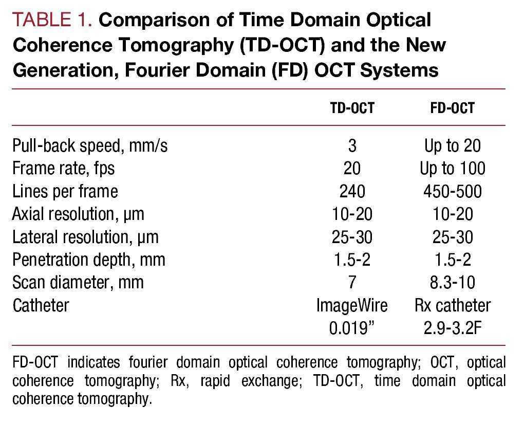
Second Generation Optical Coherence Tomography In Clinical Practice High Speed Data Acquisition Is Highly Reproducible In Patients Undergoing Percutaneous Coronary Intervention Revista Espanola De Cardiologia English Edition
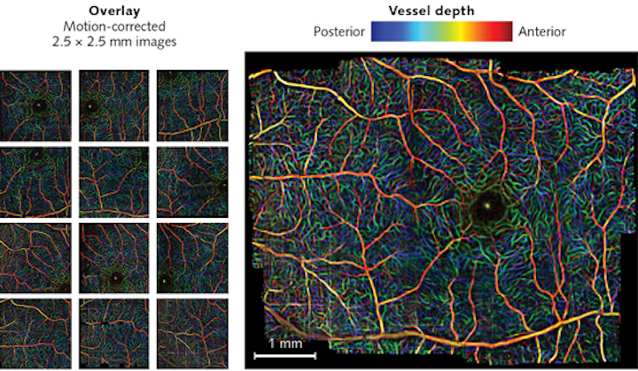
Optical Coherence Tomography Advances In Functional Oct Laser Focus World
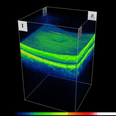
Optical Coherence Tomography Oct Lawrence And Harris
1
Bol Com Optical Coherence Tomography In Glaucoma Boeken

Optical Coherence Tomography Oct Nkt Photonics

Optical Coherence Tomography Vulnerable Plaque
Optical Coherence Tomography Oct Macula

Zeiss Oct Systems Oct Solutions Designed For The Way You Work Zeiss Medical Technology Zeiss International
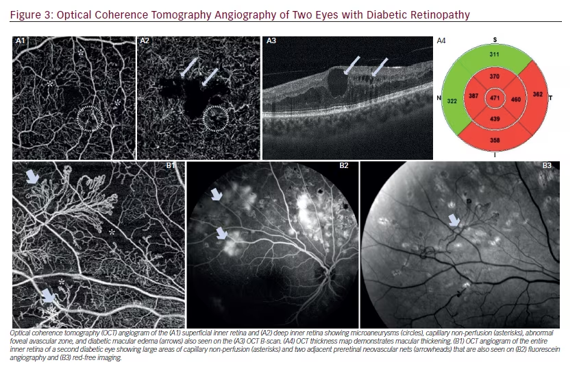
Advances In Optical Coherence Tomography Angiography Touchophthalmology
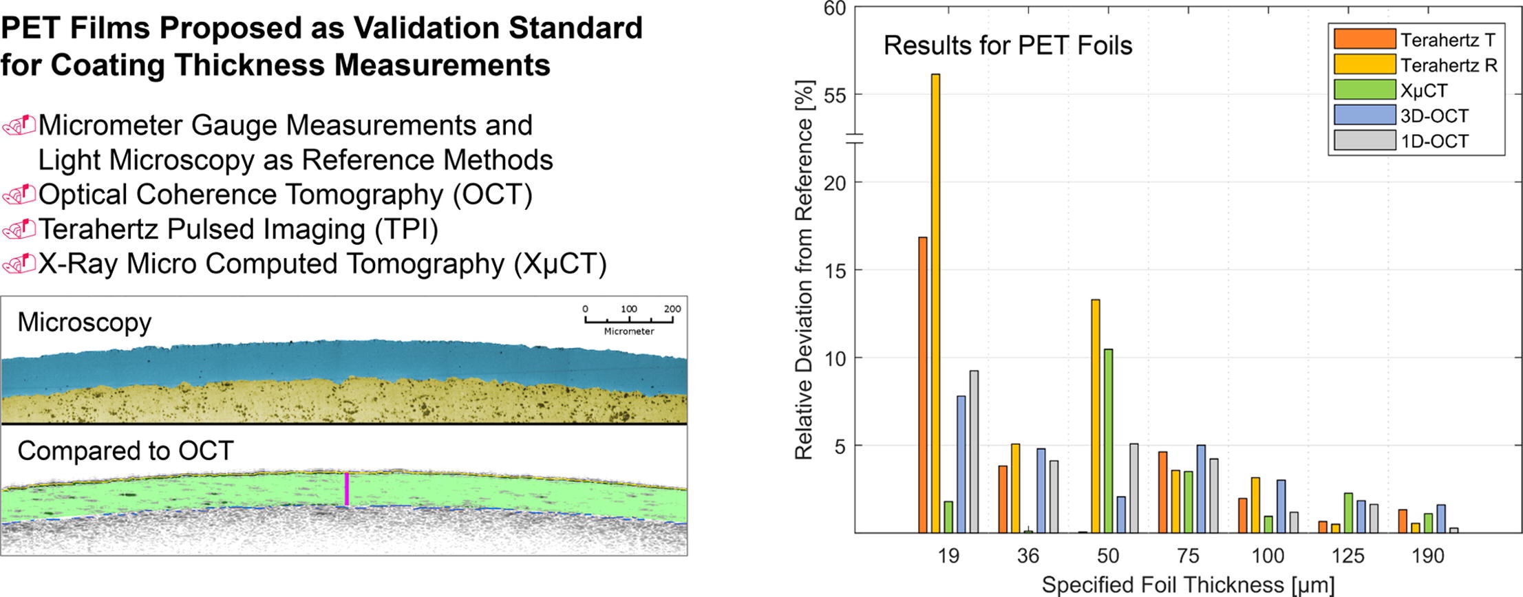
At Line Validation Of Optical Coherence Tomography As In Line At Line Coating Thickness Measurement Method Pharma Excipients
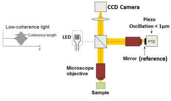
Optical Coherence Tomography Wikipedia
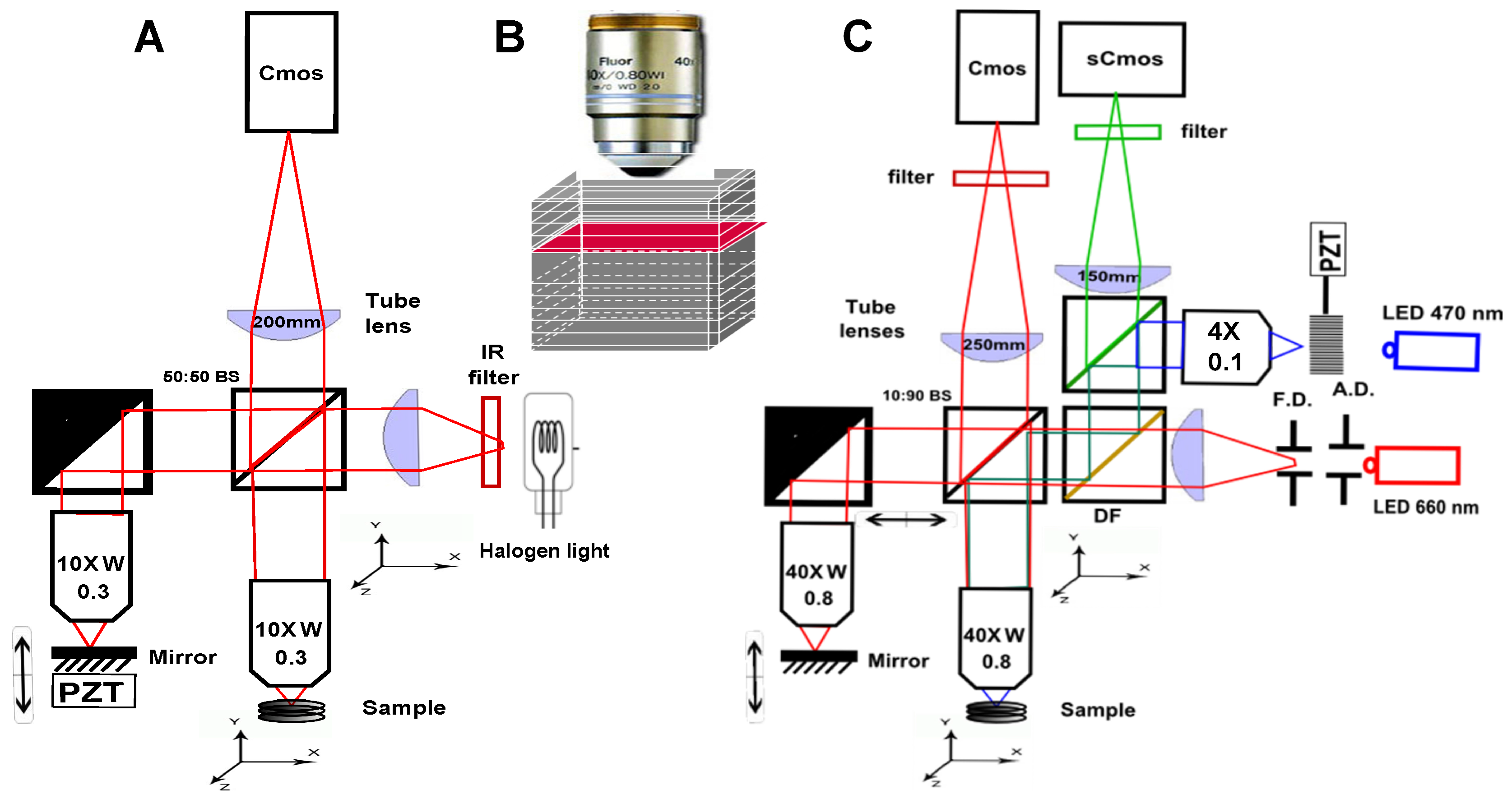
Applied Sciences Free Full Text Full Field Optical Coherence Tomography As A Diagnosis Tool Recent Progress With Multimodal Imaging Html
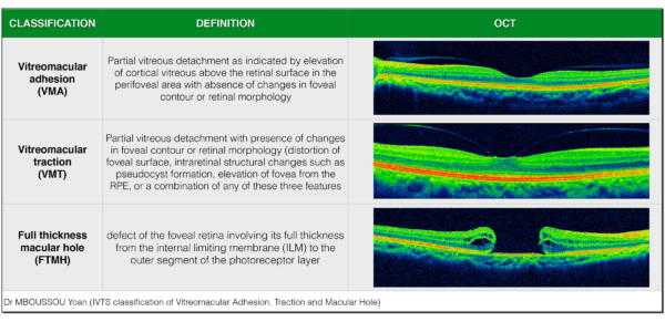
Diagnosing Eye Conditions With Optical Coherence Tomography Flei

Optical Coherence Tomography Biophotonics Imaging Laboratory

Diagnostic Accuracy Of Wide Field Map From Swept Source Optical Coherence Tomography For Primary Open Angle Glaucoma In Myopic Eyes American Journal Of Ophthalmology

Global Optical Coherence Tomography Market 17 21 Conventional Oct Systems Dominates The Global Market Technavio Business Wire

Dynamic Full Field Optical Coherence Tomography 3d Live Imaging Of Retinal Organoids Eurekalert Science News
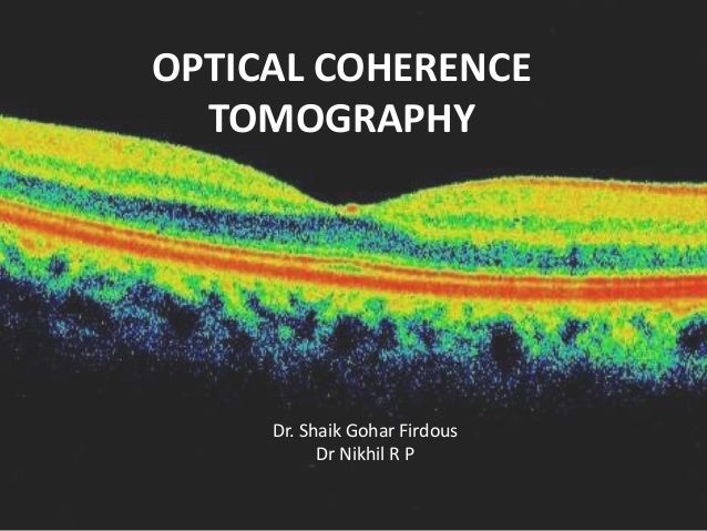
Oct
Research Vu Nl Files Chapter 2 principles of oct Pdf

Clinical Translation Of Handheld Optical Coherence Tomography Practical Considerations And Recent Advancements
Oct Tutorial Wasatch Photonics
Past And Present Terminology For The Retinal And Choroidal Structures In Optical Coherence Tomography Touchophthalmology

Optical Coherence Tomography Oct Longer Wavelengths Can Improve Imaging Depths
Optical Coherence Tomography Luna

Latest Trends In Optical Coherence Tomography Oct Voler Systems
Osa Common Path Phase Sensitive Optical Coherence Tomography Provides Enhanced Phase Stability And Detection Sensitivity For Dynamic Elastography

Optical Coherence Tomography Angiography An Overview Of The Technology And An Assessment Of Applications For Clinical Research British Journal Of Ophthalmology
Optical Coherence Tomography And Color Fundus Photography In The Screening Of Age Related Macular Degeneration A Comparative Population Based Study
Optical Coherence Tomography Oct Ibsen Photonics

Optical Coherence Tomography A Guide To Interpretation Of Common Macular Diseases Bhende M Shetty S Parthasarathy Mk Ramya S Indian J Ophthalmol
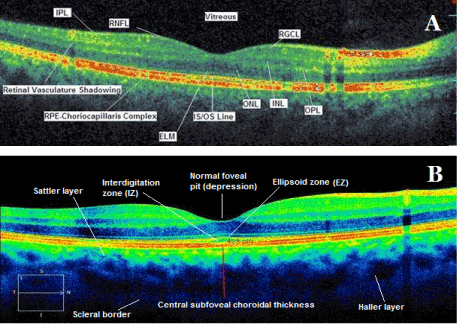
The New Landmarks Findings And Signs In Optical Coherence Tomography
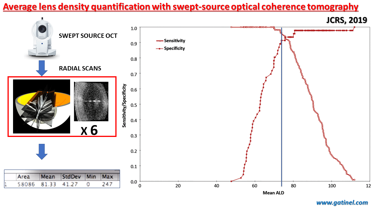
Average Lens Density Quantification With Swept Source Optical Coherence Tomography Optimized Automated Cataract Grading Technique Panthier C De Wazieres A Rouger H Moran S Saad A Gatinel D Jcrs December 19 Docteur
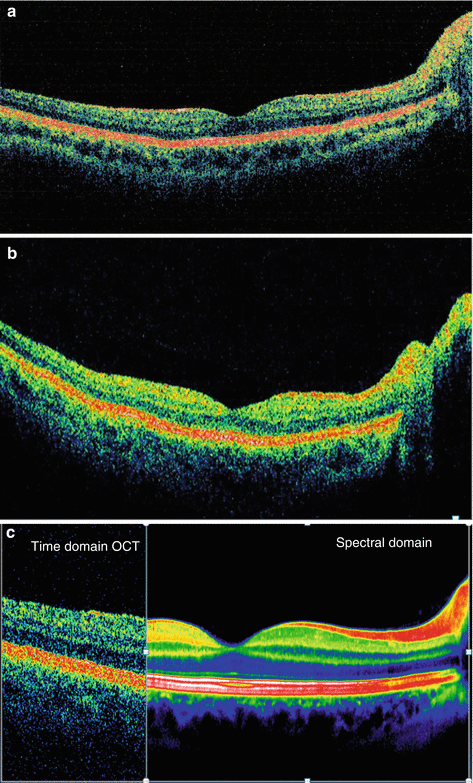
Optical Coherence Tomography As A Biomarker In Clinical Trials For Ophthalmology And Neurology Radiology Key

Optical Coherence Tomography Eyewiki

Optical Coherence Tomography Basic Explanation Youtube

Optical Coherence Tomography Angiography Atlas A Case Study Approach Slack Books
Osa En Face Optical Coherence Tomography A Technology Review Invited

Introduction To Optical Coherence Tomography Oct Background Optical Coherence Tomography Oct 1 2 Is An Imaging Technique Which Works Similar To Ultrasound Simply Using Light Waves Instead Of Sound Waves By Using The Time Delay Information
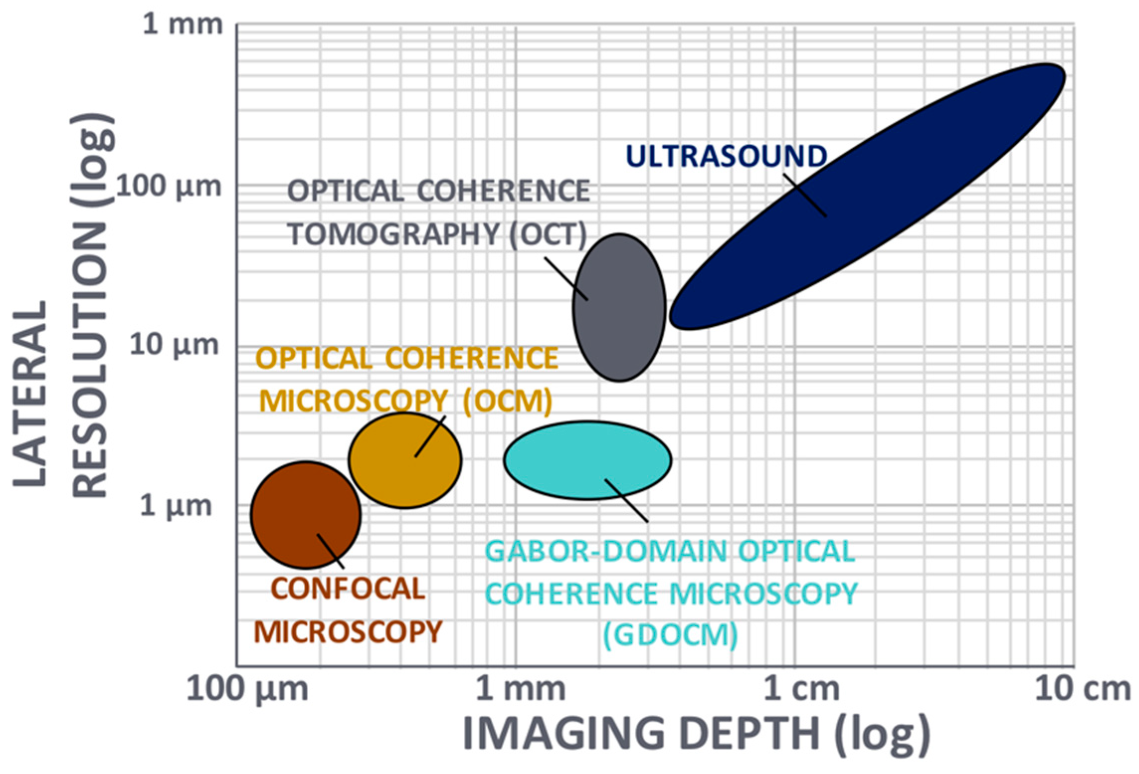
Applied Sciences Free Full Text Ten Years Of Gabor Domain Optical Coherence Microscopy

Optical Coherence Tomography Oct Fundus Image Of Wet Age Related Download Scientific Diagram

Optical Coherence Tomography Oct In Barcelona Ico

Reconstruction And Spectral Analysis For Optical Coherence Tomography File Exchange Matlab Central
Optical Coherence Tomography Market Size Report 24

Images Of Time Domain Optical Coherence Tomography Top Left Spectral Download Scientific Diagram
Frontiers Multimodal Optical Medical Imaging Concepts Based On Optical Coherence Tomography Physics
Optical Coherence Tomography Oct Principle And Technical Realization Springerlink

The Next Innovation In Pci Is Not A Stent The Value Of Optical Coherence Tomography Oct Cath Lab Digest

A Raw Optical Coherence Tomography Oct Data Consist Of 1024 Download Scientific Diagram
Optical Coherence Tomography Oct Market Size Share

Accuracy Of Optical Coherence Tomography For Diagnosing Glaucoma An Overview Of Systematic Reviews British Journal Of Ophthalmology

Enhanced Imaging Of The Esophagus Optical Coherence Tomography Sciencedirect

Global Optical Coherence Tomography Market 17 21 Conventional Oct Systems Dominates The Global Market Technavio Business Wire

Bol Com Handbook Of Retinal Oct Optical Coherence Tomography Jay Duker Boeken

Optical Coherence Tomography Oct Longer Wavelengths Can Improve Imaging Depths

Three Dimensional Multifunctional Optical Coherence Tomography For Skin Imaging

Optic Coherence Tomography

Detection Of Glaucomatous Optic Neuropathy With Spectral Domain Optical Coherence Tomography A Retrospective Training And Validation Deep Learning Analysis The Lancet Digital Health

Optical Coherence Tomography Wikipedia

Enhanced Imaging Of The Esophagus Optical Coherence Tomography Sciencedirect

Optical Coherence Tomography Oct
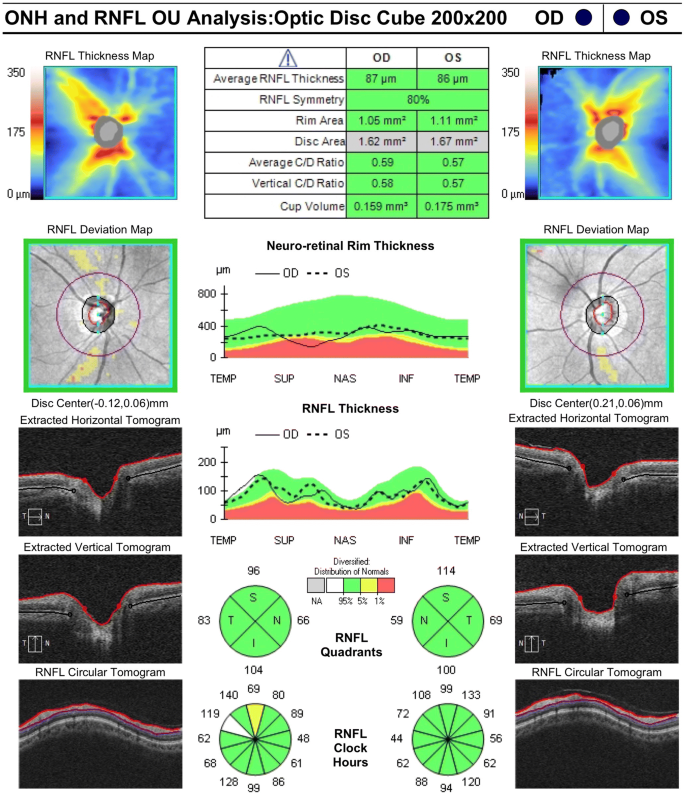
Diversity In Optical Coherence Tomography Normative Databases Moving Beyond Race International Journal Of Retina And Vitreous Full Text
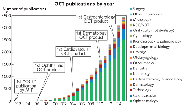
Optical Coherence Tomography Beyond Better Clinical Care Oct S Economic Impact Laser Focus World
Longitudinal Optical Coherence Tomography Oct Of Lasered Mouse Download Scientific Diagram

Predicting The Glaucomatous Central 10 Degree Visual Field From Optical Coherence Tomography Using Deep Learning And Tensor Regression American Journal Of Ophthalmology


